In July 2020, he was readmitted to our
hospital with complaints of headache, vomiting, and
facial paralysis. Peripheral blood examination
showed HB of 13 g/dl, WBC of 14000/mm3, PLT of
3,30,000/mm3. Bone marrow examination showed no
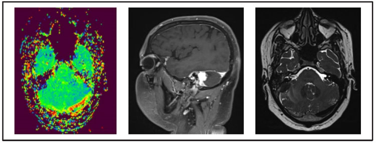
evidence of leukemia cells. Contrast-enhanced
computed tomography (CECT) brain showed
temporal bone-soft tissue thickening surrounding the
facial nerve canal and soft tissue opacity in the right
external auditory canal. Magnetic resonance images
and spectroscopy (MRI and MRS) of the brain suggest
altered signal intensity lesion along the sigmoid sinus
on the right side, infiltrate the right cerebellar
hemisphere with minimal perilesional edema. The
lesion appears iso-intense on T1W, hypo-intense on T2W and FLAIR, and shows intense post-contrast
enhancement. These CT scan and MRI scan findings
were compatible with those of myeloid sarcoma (MS)
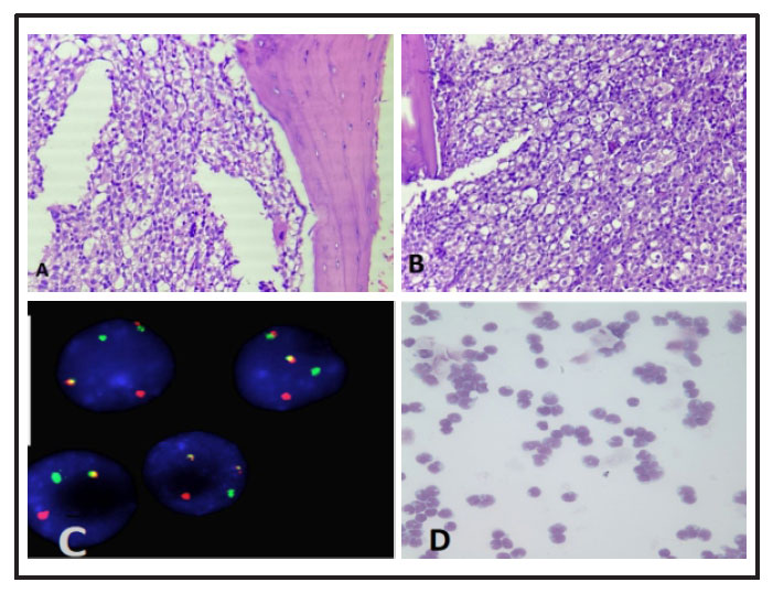
(Figure 1). CSF cytology was positive for malignant
cells, involvement by AML was suggested, and a
FISH study for t (8;21) from peripheral blood shows
the sample was positive for the AML1-ETO fusion
gene. (Figure 2)5
As there was no evidence of leukemia in the
bone marrow, an isolated recurrence of the MS of the
brain was suspected. Subsequently, he was given
biweekly triple intrathecal injections consisting of
cytarabine (70 mg), methotrexate (12 mg), and
prednisolone (50 mg) along with whole-brain
radiation (24 Gy in 12 fractions) till 19.8.2020.
After
five courses of injections with intrathecal cytarabine,
prednisolone, and methotrexate, CSF cytology of
three consecutive times showed negative for
malignancy, facial paralysis also improved.
Furthermore, he had no other neurological deficit except facial nerve palsy. Though there was no bone
marrow relapse, systemic chemotherapy, including
cytarabine 3 gm/m2 twice daily for three consecutive
days with a cumulative dose of 18 gm/m2 was
scheduled. Unfortunately, he was lost to follow up.
Recently, he presented with respiratory distress and
peripheral blood examination showed Hb of 7 gm/dl,
WBC of 58000/mm3, and platelets of 22000/mm3 with
21% blasts which suggest medullary relapse and
succumbed to the disease.
Discussion
The first case of AML with MS was described
by Turk in 1903 and suggested the origin is the same
for both the tumors.13 MS can occur in different sites
such as bones, soft tissues, skin, lymph nodes, central
nervous system, bladder, and breast.14 In the study by
Pileri et al of 92 patients with newly diagnosed MS,
35% and 38% had a simultaneous or previous treated
AML.11 The molecular and cytogenetic AML
mutations might be associated with the development
of MS. MS with translocation t(8;21)-positive cases
commonly occur in the orbital, and CNS region in
children,12 while patients with inv(16) have a high
incidence of stomach, intestine, or breast
involvement, specifically in adults.11 Our case lies in
the rare location of the MS and its relationship with an
AML with translocation t(8;21).
Byrd and Weiss 15 reviewed 24 patients from
various trials since 1973 with patients having isolated
recurrences of MS following prior AML treatment.
The isolated MS relapse generally develops bone
marrow relapse. In these patients, the mean time
interval to develop bone marrow relapse was 7
months, and the prognosis was poor. Only 3 of 24
patients had MS of the brain.16,17 The mean time
interval from diagnosis of AML to isolated MS relapse
was 2 years. All patients were treated with irradiation,
intrathecal injection, and/or operation. Systemic
chemotherapy was administered in three patients
during marrow remission.16 Six patients remained
alive even though the follow-up periods were varied.
In our study, the time interval from diagnosis of AML
to isolated MS relapse was 13 months. The time
interval to develop bone marrow relapse was 21
months.
Gustavo et al reviewing the literature,
identified 21 cases with intracranial MS.17 Fifty-four
percent had intraparenchymal lesions of the brain,
and 45% of the patients had lesions in the extra-axial
brain compartment. MS appears even before the initial
diagnosis of AML by years in 25% of the patients.18 Of
the total patients, 91% showed a hyper-dense lesion
on a non-contrast CT scan.
Migration of leukemic cells from the bone
marrow of periosteum and dura matter into the brain
parenchyma can occur once there is disruption of the
blood-brain barrier. Bone destructions are not
commonly observed with MS. Out of 24 patients, 1
patient showed visible bone destruction of the
temporal bone and simultaneous involvement of
temporal lobe parenchyma.19 Seven patients were
reviewed with brain MRI. MS showed either a hyper,
iso-or hypo-intense signal on T2-weighted images. 4
patients showed T2 hyperintensity while 3 patients
showed T2 iso or hypo-intensity.19 In our case, MS of
the brain was diagnosed by MRI and CSF cytology.
MRI brain of our patient showed hypo-intensity on
T2-weighted images.
The currently recommended treatment
options for MS are the combination of chemotherapy
and radiotherapy. There are no pathologic or clinical
prognostic features, however, survival is better in
patients who undergo allogenic bone marrow
transplant.11 Tsimberidou et al assessed the outcome of
23 patients with AML was compared with MS, and
they found that the event-free survival was longer in
patients with isolated MS.20
Recently, Lee et al in a meta-analysis of 82
studies in which variables such as the extent of
resection, treatment modality, and mortality were
correlated and they found that surgical resection and
extent of resection were not significantly associated
with mortality. Patients who received chemotherapy
or radiotherapy had lower rates of mortality versus
patients who did not received chemotherapy or
radiotherapy.21 In the present case study the patient
received whole-brain radiation therapy with biweekly
intrathecal chemotherapy. After these treatments, CSF
cytology of three consecutive times showed no
evidence of malignancy, and there was an
improvement in facial paralysis. Even though there
was no relapse in the bone marrow, prophylactic
chemotherapy was planned. Unfortunately, he was
lost to follow up. Recently, he presented with
respiratory distress and peripheral blood examination
showed Hb of 7 gm/dl, WBC of 58000/mm3, and
platelets of 22000/mm3 with 21% blasts which
suggest medullary relapse and succumbed to the
disease. From these findings, we advise that
chemotherapy must be started after the completion of
local therapy of the brain.
Conclusion
Since randomized prospective studies are
lacking, there is no proper consensus on the treatment
of MS. The currently recommended treatment
regimen in patients presenting with isolated MS of the
brain is localized treatment with cranial irradiation
and or operation with intrathecal chemotherapy followed by prophylactic systemic chemotherapy. As
from these findings, we advise that chemotherapy
should be started after the completion of local
treatment to the brain. Close follow-up of the patient is
needed following AML treatment and any new onset
of neurological symptoms should be thoroughly
evaluated.
References
1. Döhner H, Weisdorf DJ, Bloomfield CD et al:
Acute Myeloid Leukemia. N Engl J Med 2015;
373: 1136-1152
2. Weinberg OK, Sohani AR, Bhargava P et al:
Diagnostic workup of acute myeloid leukemia.
Am J Hematol 2017; 92: 317-321
3. Arber DA, Borowitz MJ, Cessna M et al: Initial
Diagnostic Workup of Acute Leukemia.
Guideline from the College of American
Pathologists and the American Society of
Hematology. Arch Pathol Lab Med 2017; 141:
1342-1393
4. Döhner H, Estey E, Grimwade D et al: Diagnosis
and management of AML in adults. 2017 ELN
recommendations from an international expert
panel. Blood 2017; 129: 424-447
5. Arber DA, Orazi A, Hasserjian R et al: The 2016
revision to the World Health Organization
classification of myeloid neoplasms and acute
leukemia. Blood 2016; 127: 2391-2405
6. Byrd JC, Edenfield WJ, Shields OJ et al:
Extramedullary myeloid cell tumors in acute
nonlymphocytic leukemia. A clinical review. J
Clin Oncol 1995; 13: 1800-1816
7. Kawamoto K, Miyoshi H, Yoshida N et
al:Clinicopathological, cytogenetic, and
prognostic analysis of 131 myeloid sarcoma
patients. Am J Surg Pathol 2016; 40: 1473-1483
8. Lai Y-Y, Qiu J-Y, Jiang B et al: Characteristics and
prognostic factors of acute myeloid leukemia with
t(8;21) (q22; q22) Zhongguo Shi Yan Xue Ye Xue
Za Zhi. 2005; 13: 733-740
9. Reinhardt D, Creutzig U: Isolated myelosarcoma
in children-update and review. Leuk Lymphoma
2002; 43: 565-574
10. Stve HK, Sandahl JD, Abrahamsson J et al:
Extramedullary leukemia in children with acute
myeloid leukemia: A population-based cohort
study from the Nordic Society of Pediatric
Hematology and Oncology. Pediatr Blood Cancer
2017; 64: e26520
11. Pileri SA, Ascani S, Cox MC et al: Myeloid
sarcoma: Clinico-pathologic, phenotypic, and
cytogenetic analysis of 92 adult patients.
Leukemia 2007; 21: 340-350
12. Bönig H, Göbel U, Nürnberger et al: Bilateral
exophthalmus due to retro-orbital chloromas in a
boy with t(8;21)-positive acute myeloblastic
leukemia. Pediatric Hematology and Oncology
2002; 19 :597-600
13. Rappaport H: Tumors of the Hematopoietic
System, Atlas of Tumor Pathology. Washington,
DC, USA: Armed Forces Institutes of Pathology;
1966
14. Mangla D, Dewan M, Meyer DR. Adult orbital
myeloid sarcoma (granulocytic sarcoma): two
cases and review of the literature. Orbit 2012;
31:438-440
15. Byrd JC, Weiss RB: Recurrent granulocytic
sarcoma: an unusual variation of acute
myelogenous leukemia associated with 8;21
chromosomal translocation and blast expression
of the neural cell adhesion molecule. Cancer
1994; 73: 2107-2712
16. Infante AJ, MacRae MA, Forbes GS et al:
Intracranial granulocytic sarcoma complicating
childhood acute myelomonocytic leukemia. Am J
Pediatr Hematol Oncol 1981; 3: 173-176
17. Gustavo M, Cayci Z: Intracranial CNS
manifestations of myeloid sarcoma in patients
with acute myeloid leukemia: Review of the
literature and three case reports from the author’s
Institution. J Clin Med 2015; 4: 1102-1112
18. Avni B, Koren-Michowitz M: Myeloid sarcoma.
Current approach and therapeutic options. Adv
Hematol 2011; 2: 309-316
19. Murakami M, Uno T, Nakaguchi H et al: Isolated
recurrence of intracranial and temporal bone
myeloid sarcoma-Case report. Neurol Med Chir
Tokyo 2011; 51: 850-854
20. Tsimberidou AM, Kantarjian HM, Wen S et al:
Myeloid sarcoma is associated with superior
event-free survival and overall survival compared
with acute myeloid leukemia. Cancer 2008; 113:
1370-1378
21. Lee D, Omofoye OA, Nuño MA et al: Treatment
Outcomes of Intracranial Myeloid Sarcomas: A
Meta-Analysis. World Neurosurgery 2021; 148:
29-37