Gujarat Cancer Society Research Journal
 

Case Report
Steroid Cell Tumour Ovary: An Unusual Presentation

Abdul Shukur Remi(1), Patel Bijal M(2), Dave Pariseema S(3), Parekh Chetana(4), Kar Bijoy(5)
Resident(1), Professor(2), Professor and Head(3), Associate Professor(4), Lecturer(5)
Department of Gynaecological Oncology, The Gujarat Cancer and Research Institute, Asarwa, Ahmedabad, Gujarat, India.
Corresponding Author: bijal.patel@gcriindia.org


(1)https://orcid.org/0000-0001-7212-3040
(2)https://orcid.org/0000-0002-5446-1959
(3)https://orcid.org/0000-0003-3300-4414
(4)https://orcid.org/0000-0003-4811-5889
(5)https://orcid.org/0000-0001-6176-8687

Volume : 23 / Number : 1 / April 2021


Summary
Steroid cell tumours of the ovary are extremely rare sex hormone secreting tumours with an incidence of only 0.1% of all ovarian tumours. We report a case of a steroid cell tumour ovary in a 68-year-old postmenopausal women who presented with
postmenopausal bleeding. Her examination was unremarkable except for bulky uterus. CT scan showed a 5.3 x 4.5 cm left adnexal solid cystic lesion with increased endometrial thickness. She underwent primary staging laparotomy with frozen section of left adnexal mass. Final histopathology was reported as steroid cell tumour of left ovarian mass, FIGO stage IA. She is on observation and remains recurrence free. Keywords: Steroid cell tumour, ovary

Introduction
Steroid cell tumours of the ovary are extremely rare and constitute about 0.1 % of ovarian tumours. These tumours are further divided into three subtypes according to the cell of origin into stromal luteoma, leydig cell tumour and steroid cell tumournot otherwise specified (NOS).Of these subtypes, steroid cell tumours-NOS constitute 56% of steroid cell tumours.1 These tumours have been diagnosed from early childhood to the ninth decade of life.1 They usually present with symptoms like hirsutism and virilization(56-77%) , abnormal uterine bleeding and in postmenopausal females with postmenopausal bleeding. Morphologically steroid cell tumour -NOS present as solid well-defined masses in about 89% of the cases. Rarely, they present as cystic masses, in 1.6% of cases.2 Steroid cell tumours- NOS are clinically malignant in 25-43% of cases.1 The following is a case report of steroid cell tumour of the ovary diagnosed in a 68-year-old postmenopausal female who presented with postmenopausal bleeding and hirsutism.

Case Report
A 68-year-old para 5 live 4 woman presented with complaints of postmenopausal bleeding of one month duration along with passage of clots for 3 days. On examination her uterus was bulky for age. A computerised tomography of abdomen pelvis was done which showed a 5.3 x4.5 cm left adnexal solid cystic lesion and an endometrial thickness of 10 mm. An endometrial biopsy and endocervical curettage was done which was negative for malignancy. Endometrial biopsy showed proliferative endometrium. Her CA-125, CEA and CA19-9 were normal. Inhibin B was raised - 23.5pg/ml. She underwent primary staging laparotomy with frozen section of the left salpingo-oophorectomy specimen. Frozen section was reported as either sex cord stromal tumour or leutenised granulosa cell tumour. Hence proceeded with total abdominal hysterectomy and right saplingo oophorectomy, infra colic omentectomy and left pelvic lymph node dissection. IHC was done on the specimen which was positive for Vimentin, Inhibin and MIB. Final histopathology was reported as steroid cell tumour-NOS FIGO stage IA. (Figure 1 and 2) Pathological correlates for malignant behaviour included mitotic figures more than 7 per 10 HPF, presence of necrosis and size of tumor 7 cm in our case. After tumor board discussion, which included the medical oncologist, gynaec oncologist and oncopathologist, decision for keeping the patient on close observation with clinical examination and ultrasonography abdomen and pelvis was taken. Patient is on observation and remains recurrence free for the past seven months.

Figure 1: Low power view shows tumour mass clearly separated
from normal ovarian cortex (hematoxylin and eosin stain, original magnification x 100)
Figure 2:High power shows variable sized round to polygonal
tumour cells with abundant vacuolated cytoplasm and round
nuclei with prominent nucleoli (hematoxylin and eosin stain
original magnification x400)

Discussion

Steroid cell tumour ovary was first described by Scully.1 Formerly they were referred to as lipid cell tumours of the ovary. The incidence of a steroid cell tumours is less than 0.1% of all ovarian tumours.1 Most commonly they present in the childbearing age group or third and fourth decade of life and very rarely do they occur in postmenopausal women. These tumours are hormone secreting and hence cause androgenic manifestation.

These tumours generally present with symptoms of virilisation and menstrual irregularity and hence patients with such symptoms should be suspected of having adrenal and ovarian tumour which should be ruled out clinically. In cases of rapid onset hirsuitism and virilisation, serum testosterone value above 200 ng/dL is important in diagnosing neoplastic source of hirsuitism. It is also useful in post treatment follow up of patients if initially elevated. Our patient had a proliferative endometrium on endometrial biopsy. This along with her history of post-menopausal bleeding indicates an oestrogen secreting tumour. Hyperestrogenemia presenting as menorrhagia or post-menopausal bleeding has been reported in 6 to 23% of women.1 In most of the cases, the diagnosis of steroid cell tumours- NOS is made post operatively.

Majority of steroid cell tumours-NOS are unilateral, solid and well circumscribed with size ranging from 1.2 to 45 cms.1 Grossly a combination of solid cystic tumours have also been reported however purely cystic tumours are extremely rare. Cut surface range from yellow to orange to red or brown 
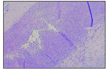
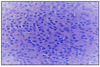
depending on the lipid content.1,2 Microscopically the tumour cells are polygonal and have abundant cytoplasm that ranges from eosinophilic (lipid-poor) to pale and vacuolated (lipid-rich), arranged in sheets with prominent central nucleus and centrally placed round nuclei. Immunohistochemistry for inhibin, calretin and melan A are sensitive markers for steroid cell tumours- NOS.3

The tumour in our case was solid cystic mass of 7x6 cm, with cut surface showing yellow colour with areas of necrosis and haemorrhage. Clinico pathologic correlation is essential for management of these cases. Treatment of these tumours are based on histological picture, surgical staging and patients desire to preserve fertility. As our patient was postmenopausal, we did a complete staging surgery.

Clinico pathologic parameters which correlate with malignant behaviour of these tumours include advanced age at the time of presentation, size of tumour of 7 cm or more (78%), mitotic figures more than 2 / 10 hpf (92%), grade 2 to 3 nuclear atypia (64%), presence of necrosis (86%) and haemorrhage (77%). 1,4

In the present case even though the patient had adverse prognostic factors like older age at presentation, increased mitotic rate, size of the tumour of 7 cm and presence of necrosis and haemorrhage, the patient has been kept on close observation as she has undergone an optimal staging surgery and FIGO stage of tumour being IA. Patient is disease free till date.

Conclusion

Steroid cell tumours-NOS are very rare ovarian sex cord stromal tumours which usually present with varied symptoms like menstrual irregularities, hirsutism and abdominal pain. In postmenopausal women therapeutic complete surgery should be performed. Clinical correlation along with histopathologic examination is the gold standard that can confirm the diagnosis in most cases and in atypical cases immunohistochemistry plays a very significant role.

References

1. Hayes MC, Scully RE: Ovarian steroid cell t u m o u r s ( n o t o t h e r w i s e s p e c i f i e d ) . A clinicopathological analysis of 63 cases. Am J Surg Pathol 1987;11:835-845
2. Outwater EK, Wagner BJ, Mannion C et al: Sex Cord-stromal and steroid cell tumours of the ovary. Radio graphics 1998;18:1523-1546
3. Kurman RJ: International Agency for Research on Cancer, and World Health Organization, WHO classification of tumours of female reproductive organs, International Agency for Research on Cancer, Lyon, 4th edition, 2014, World Health Organization classification of tumours.
4. RubidoValle CD, Fuentes JL, Martinez CS et al: Ovarian steroid cell tumour associated to endometrial hyperplasia and presenting as post menopausal vaginal bleeding. Gynecol Obstet 2015;5:316
5. Sawathiparnich P, Sitthinamsuwan P, Sanpakit K: Cushing’s syndrome caused by ACTH- producing ovarian steroid cell tumour, NOS, in a prepubertal girl. Endocrine 2009;35:132-135

OPD Information

NABH Certified

Ethics Committee Accredited for NABH

ISO 15189:2012
@ The Gujarat Cancer & Research Institute, All Rights Reserved.