Gujarat Cancer Society Research Journal
 

Case Report
Mixed Endometrial Carcinoma Rare Histological Type

Gupta Rubi(1), Parekh Chetana(2), Dave Pariseema S(3), Patel Bijal M(4), Kar Bijoy(5)
Resident(1), Associate Professor(2), Professor and Head of Unit(3), Professor(4), Lecturer(5)
Department of Gynaecological Oncology
The Gujarat Cancer & Research Institute, Asarwa Ahmedabad Gujarat
Corresponding Author: chetna.parekh@gcriindia.org


(2)https://orcid.org/0000-0003-4811-5889
(3)https://orcid.org/0000-0003-3300-4414
(4)https://orcid.org/0000-0002-5446-1959
(5)https://orcid.org/0000-0001-6176-8687

Volume : 23 / Number : 1 / April 2021


Summary
WHO defines Mixed Endometrial Carcinoma (MEC) as a tumor composed of “two or more different histological types of endometrial carcinoma, at least one of which is of the type II category” The type II refers to non-endometroid (serous carcinoma or clear cell carcinoma) carcinoma. MEC comprises of about 5% of all endometrial cancers (EC), due to its rarity precise definition and exact data on occurrence is difficult to generate. Here we are reporting a case of sixty-one-year-old women presented with post-menopausal bleeding. Histopathology of endometrium revealed endometroid and clear cell components, consistent with mixed endometrial carcinoma. Patient underwent comprehensive staging laparotomy followed by adjuvant treatment with chemo-radiation. Patient is on close follow-up till date and continues to be disease free.
Keywords: Mixed Endometrial Carcinoma, Clear Cell Carcinoma, Radiotherapy, Chemotherapy

Introduction
Mixed endometrial carcinoma is composed of two or more distinct histologies in the same specimen and both the cell types must be recognisable distinctly on haematoxylin and eosin-stained pathological sections.1 According to WHO 2014 the minimum percentage of second component is arbitrarily set as 5 and can be confirmed on immunohistochemistry (IHC).1 MEC are almost clonal rather than being collision.2 Most common combination of MEC is the endometrioid with serous (endometroid with type II). Quantification of each component is very challenging and important in respect to management and prognosis.3 Incidence of Mixed endometrial carcinoma is rarity and is reported to be less than 5%. The report below gives the time line of events of one such case..

Case Report
Sixty one year old postmenopausal, nulliparous women presented to our gynaecological oncology department (Gujarat Cancer and Research Institute) with a complaint of vaginal bleeding for one month. Her bleeding was sudden in onset with minimal flow and was not associated with passage of clots. She was postmenopausal for 12 years. She was known case of hypertension and diabetes. She had no history suggestive of cancer in the family. On physical examination, she was overweight with body mass index 28.6 Kg/m2 On per-speculum examination cervical was pinpoint, looks healthy and vagina was normal. On bimanual examination uterus was bulky retroverted, bilateral fornixes were free.

Her sonography revealed 44x49 mm heterogenous echotexture lesion in left lateral wall of uterus, with internal vascularity and loss of endomyometrial junction. Lesion had myometrial invasion of more than 50%. Patient underwent dilatation and curettage (D & C). Histopathology of D&C material revealed endometroid carcinoma with clear cell change. IHC was positive for napsin, CK 7, ER, vimentin and negative for CK 20, PR, P40, CEA that indicated endometroid carcinoma with clear cell change. Further imaging with MRI was done to determine extent of disease. MRI revealed 33x53x35 mm altered intensity lesion in endometrium with endocervical gland involvement. Lesion invades more than half of myometrium. Few sub centimetric lymph nodes were noted along bilateral iliac vessels. Her baseline blood counts, liver and kidney function were with in normal range and her CA-125 was 22.6 IU/ml. Patient was diagnosed as carcinoma endometrium and underwent staging laparotomy with peritoneal cytology, total abdominal hysterectomy, bilateral salpoingo-ophorectomy, bilateral pelvic
lymph node dissection, infra colic omentectomy. Final histopathology revealed tumor size 5 X 5 cm, mixed histology of clear cell and endometroid carcinoma. Clear cell accounting 70% of carcinoma while endometroid accounting to 30%. Myometrial invasion of about 80% (3/4 thickness of myometrial wall) along with lower uterine segment involvement. Uterine serosa, cervical stroma, bilateral tubes and ovaries, omentum and lymph nodes were free of tumor. IHC was positive for ER, PR confirmed endometroid nature while positivity for napsin, p53 confirmed clear cell component. Her FIGO stage was IB with high intermediate risk. After discussing the case in multidisciplinary tumor board adjuvant treatment was decided. She received radiotherapy (EBRT 50 Gy / 25# plus brachytherapy with 6.5 Gy / 2#) followed by chemotherapy (paclitaxel and carboplatin 4 cycle 3 weekly). Patient is on regular follow up and disease free till date.

Figure 1: Top right corner shows malignant endometroid
carcinoma and left lower shows clear cell component on H & E
stain.
Figure 2: IHC showing napsin cytoplasmic positivity in clear ell
(left) and ER & PR positivity in endometroid (right) component.

Discussion

Endometroid adenocarcinoma (type I) is the most common histologic subtype accounting 80-85% of all EC. While clear cell carcinoma (type II) accounting for only 1-5%. Type II EC occurs in older population and associated with aggressive clinical behaviour and had poor prognosis compared to type I EC. MEC is a rare histological variant composed of type I and type II EC. It is not considered as a morphological variant of endometrioid cancer that stimulate type II component. WHO mandates IHC staining to confirm subtype of type II and any amount of type II component in endometrioid carcinoma qualified as MEC. It has aggressive behaviour and poor outcome compared to endometroid carcinoma (type I) and similar to pure serous or clear cell carcinoma of endometrium (Type II). Kaban et al in 2018 also proposed similar result that, MEC have same prognosis and risk of metastases as patients with pure endometrial serous carcinoma.4

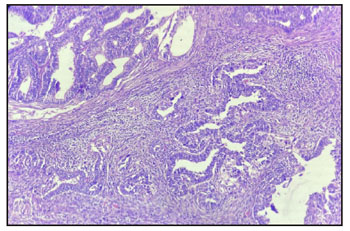
Diagnosis of MEC is diagnosis of exclusion. A pathologic morphology on H & E stain is insufficient to diagnose and it mandates the 
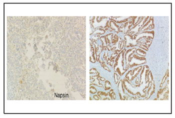
conformation of the mixed nature by IHC. A Combination of ER, PR, p53 and napsin are used to distinguish type I from type II EC. A positivity of ER, PR and a negativity of napsin favours endometroid subtype whereas napsin positivity favours the diagnosis of clear cell subtype. In endometroid subtype p53 is almost always negative, where as it may rarely be positive in clear cell subtypes as opposite to its high positivity in serous histologies. MEC is consider as high grade regardless of the amount of type II component in it.5 Wenhui et al in 2019 reported that any amount of non-endometroid component in MEC indicate poor prognosis and warrant rigorous adjuvant treatment and close follow up. They also reported better survival in MEC with aggressive treatment compared to pure non endometroid carcinoma.6

Treatment plan should be made, considering the aggressive counterpart in MEC as planned in our case. Comprehensive surgical staging is cornerstone of management in type II EC. These patients often experience local, nodal and distant recurrence. According to Postoperative Radiation Therapy in Endometrial carcinoma (PORTEC-3) trial high risk endometroid and early-stage clear cell carcinoma should be treated with chemo-radiation. Similarly in our case patient also received comprehensive surgical
s t a g i n g f o l l o w e d b y E B RT a n d a d j u v a n t chemotherapy considering aggressive nature of clear cell carcinoma of EC even in stage IB for better survival.

Conclusion

As MEC has inferior survival outcome and high chance of metastasis compared to endometroid adeno carcinoma they need rigorous adjuvant treatment and follow up.

References

1. Zaino R, Carinelli S, Ellenson LH et al: WHO classification of tumours of the uterine corpus. In: Epithelial tumours and precursors: mixed carcinoma 2014; 132
2. Köbel M, Meng B, Hoang LN et al: Molecular analysis of mixed endometrial carcinomas shows clonality in most cases. Am J Surg Pathol 2016; 40:166-180
3. Quddus MR, Sung CJ, Zhang C, Lawrence WD et al: Minor serous and clear cell components adversely affect prognosis in ''mixed-type'' endometrial carcinomas: a clinicopathologic study of 36 stage-I cases 2010;17:673-678
4. Alpaslan Kaban, Samet Topuz, Hamdullah Sözen et al: Clinicopathologic and survival results in serous endometrium carcinoma and subgroup analysis for mixed serous and pure serous histology. J Turk Ger Gynecol Assoc 2018; 19:23- 28
5. Rabban JT, Gilks CB, Malpica A et al: Issues in the differential diagnosis of uterine low-grade endometrioid carcinoma, including mixed endometrial carcinomas: Recommendations from The International Society of Gynecological Pathologists. Int J Gynecol Pathol 2019; 38: S25- 39
6. Wenhui Li, Lei Li, Ming Wu, Jinghe Lang, et al: The prognosis of stage IA mixed endometrial carcinoma. Am J Clin Pathol 2019; 152:616-624

OPD Information

NABH Certified

Ethics Committee Accredited for NABH

ISO 15189:2012
@ The Gujarat Cancer & Research Institute, All Rights Reserved.